Ultrasounds are high frequency waves. Ultrasounds are able to travel along well defined paths even in the presence of obstacles. Ultrasounds are used extensively in industries and for medical purposes.
Ultrasound is generally used to clean parts located in hard-to-reach places, for example, spiral tube, odd shaped parts, electronic components etc.
Objects to be cleaned are placed in a cleaning solution and ultrasonic waves are sent into the solution. Due to the high frequency, the particles of
dust, grease and dirt get detached and drop out. The objects thus get thoroughly cleaned.

defective locations inside a metal block.
Ultrasounds can be used to detect cracks and flaws in metal blocks. Metallic components are generally used in construction of big structures like buildings, bridges, machines and also scientific equipment. The cracks or holes inside the metal blocks, which are invisible from outside reduces the strength of the structure. Ultrasonic waves are allowed to pass through the metal block and detectors are used to detect the transmitted waves. If there is even a small defect, the ultrasound gets reflected back indicating the presence of the flaw or defect.
Ordinary sound of longer wavelengths cannot be used for such purpose as it will bend around the corners of the defective location and enter the detector.
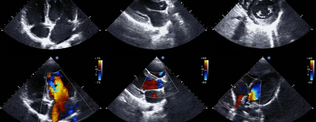
Ultrasonic waves are made to reflect from various parts of the heart and form the image of the heart. This technique is called ‘echocardiography’

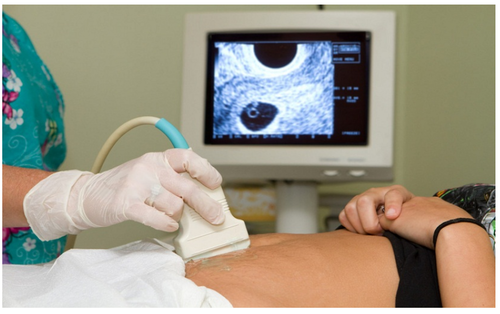
Ultrasound scanner is an instrument which uses ultrasonic waves for getting images of internal organs of the human body. A doctor may image
the patient’s organs such as the liver, gall bladder, uterus, kidney, etc. It helps the doctor to detect abnormalities, such as stones in the gall bladder and kidney or tumours In different organs. In this technique the ultrasonic waves travel through the tissues of the body and get reflected from a region where there is a change of tissue density. These waves are then converted into electrical signals that are used to generate images of the organ. These images are then displayed on a monitor or printed on a film. This technique is called ‘ultrasonography’.
Ultrasonography is also used for examination of the foetus during pregnancy to detect congenial defects and growth abnormalities.

Ultrasound may be employed to break small ‘stones’ formed in the kidneys into fine grains. These grains later get flushed out with urine.

