The eye is a crucial and among the most intricate sense organs that we have as humans. It aids in our ability to visualise objects as well as our perception of light, color, and depth.
Structure of Human Eye

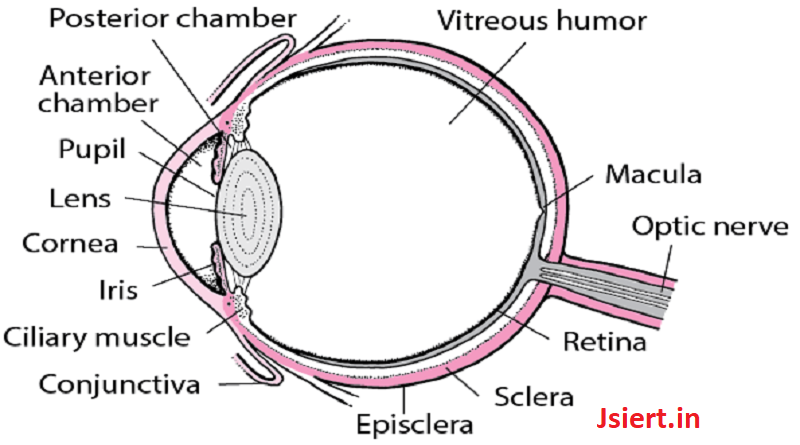
Sclera: It is the outer covering, a protective tough white layer called the sclera (white part of the eye).
Cornea: The front transparent part of the sclera is called the cornea. Light enters the eye through the cornea.
Iris: A dark muscular tissue and ring-like structure behind the cornea known as the iris. The color of the iris indicates the color of the eye. The iris also helps regulate or adjust exposure by adjusting the iris.
Pupil: A small opening in the iris is known as a pupil. Its size is controlled with the help of the iris. It controls the amount of light that enters the eye.
Lens: Behind the pupil, there is a transparent structure called a lens. By the action of ciliary muscles, it changes its shape to focus light on the retina. It becomes thinner to focus on distant objects and becomes thicker to focus on nearby objects.
Retina: It is a light-sensitive layer that consists of numerous nerve cells. It converts images formed by the lens into electrical impulses. These electrical impulses are then transmitted to the brain through the optic nerves.
Optic nerves: Optic nerves are of two types. These include cones and rods.
Cones: The nerve cells that are more sensitive to strong light are known as cones. They support colour and fine detail vision.
Rods: More sensitive to low light levels are rods, which are the optic nerve cells. Their peripheral vision is improved.
There are no sensory nerve cells at the junction of the optic nerve and retina. Therefore, the area is known as a blind spot and no vision is possible there.
There are six muscles in an eye as well. The inferior rectus, superior rectus, inferior rectus, inferior oblique, and superior oblique are all included. These muscles’ primary purpose is to provide various tensions and torques that help to further govern eye movement.
The Functions of the Human Eye

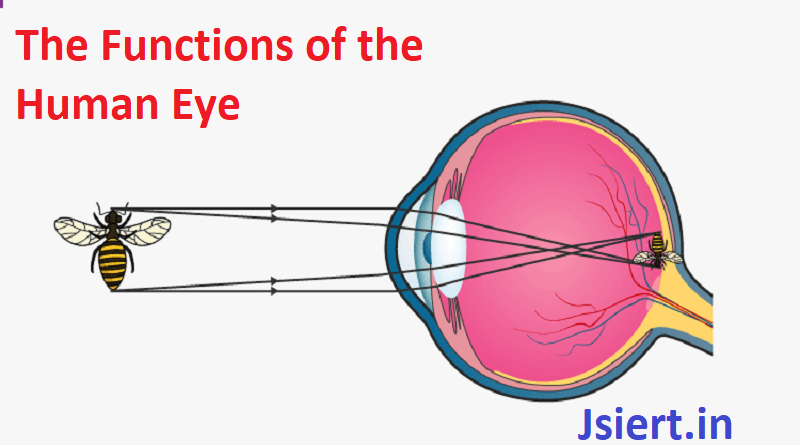
The human eye is like a camera, as we previously stated. The human eye similarly is focused and lets in light to produce images to the electronic device. In other words, light rays that are bent or refracted by distant objects pass through the cornea, crystalline lens, aqueous humor, lens, and vitreous humour before landing on the retina.
However, the idea is that light beams experience refraction as they pass through the various media. Refraction, to put it simply, is nothing more than a shift in the path of light beams as they move through various materials.
The rays are bent to create an image because they have various refractive indices. Finally, the retina receives and focuses the light rays. Rods and cones, which are photoreceptor cells in the retina, are essentially sensors for light frequency and intensity. Millions of these cells also process the image that is created, relaying the signal or nerve impulses to the brain via the optic nerve. The brain corrects this phenomenon since the image created is typically inverted. This procedure is comparable to how a convex lens works.
